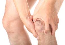
Anterior Knee Pain
Anterior knee pain is characterized by chronic pain over the front and center of the knee joint. It is common in athletes, active adolescents (especially girls) and overweight individuals.
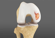
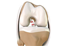
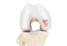
Articular Cartilage Injury
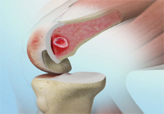
Articular or hyaline cartilage is the tissue lining the surface of the two bones in the knee joint. Cartilage helps the bones move smoothly against each other and can withstand the weight of the body during activities such as running and jumping.
Fractures of the Patella
The patella or kneecap is a small bone present in the front of your knee where the thigh bone meets the shinbone. It provides protection to your knee and attachment to muscles in the front of the thigh
Fractures of the Tibia
The lower leg is made up of two long bones called the tibia and fibula that extend between the knee and ankle. The tibia or shinbone is the larger of the two bones.
Jumper's Knee
Jumper’s knee, also known as patellar tendinitis, is inflammation of the patellar tendon that connects your kneecap (patella) to your shinbone.
Knee Arthritis
There are many conditions that can result in degeneration of the knee joint. Osteoarthritis is the most common reason that patients need to undergo knee replacement surgery.
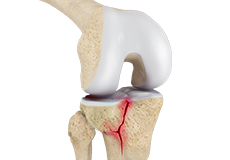
Knee Fracture
A fracture is a condition in which there is a break in the continuity of the bone. In younger individuals, these fractures are caused by high energy injuries, as from a motor vehicle accident.
Knee Infection
Knee infection is a serious medical condition that needs immediate treatment. Infection may occur followed by a knee replacement surgery or trauma and is usually caused by bacteria.
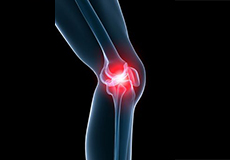
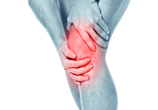
Knee Injury
If the pain and swelling are rapid, then immediate diagnosis and appropriate medical treatment are advised. Initial diagnosis includes physical and joint examination followed by an X-ray.
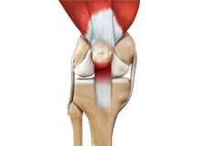
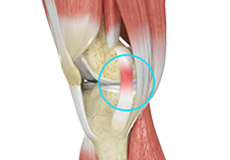
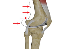
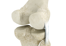
Knee Ligament Injuries
The knee is a hinge joint made up of two bones, the thighbone (femur) and shinbone (tibia). Ligaments are tough bands of tissue that connect one bone to another bone. The ligaments of the knee stabilize the knee joint. There are two important groups of ligaments that hold the bones of the knee joint together, collateral and cruciate ligaments - medial collateral ligament (MCL) and lateral collateral ligament (LCL), and anterior cruciate ligament (ACL) and posterior cruciate ligament (PCL).
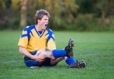
Knee Sports Injuries
Trauma is any injury caused during physical activity, motor vehicle accidents, electric shock, or other activities. Sports trauma or sports injuries refer to injuries caused while playing indoor or outdoor sports and exercising.
Knee Sprain
Knee sprain is a common injury that occurs from overstretching of the ligaments that support the knee joint. A knee sprain occurs when the knee ligaments are twisted or turned beyond its normal range, causing the ligaments to tear.
MCL Sprains
MCL sprains occur due to a sudden impact from the outside of your knee, most commonly while playing sports such as rugby and football.
Medial Meniscus Syndrome
Of the menisci within the knee, it is the medial that is more easily injured. Differences in the anatomical attachments of the medial meniscus compared to the lateral, mean that the medial meniscus becomes distorted during combined flexion and rotation movements in a manner not experienced on the lateral side.
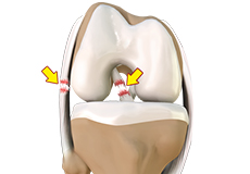
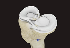
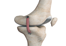
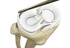
Meniscal Injuries
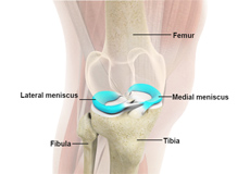
The knee is one of the most complex and largest joints in the body and is very susceptible to injury. The meniscus is a small, C-shaped piece of cartilage in the knee. Each knee consists of two menisci, medial meniscus on the inner aspect of the knee and the lateral meniscus on the outer aspect of the knee. The medial and lateral menisci act as a cushion between the thighbone (femur) and shinbone (tibia).
Multiligament Instability
The knee is a complex joint of the body that is vital for movement. The four major ligaments of the knee are anterior cruciate ligament (ACL), posterior cruciate ligament (PCL), medial collateral ligament (MCL) and lateral collateral ligament (LCL).
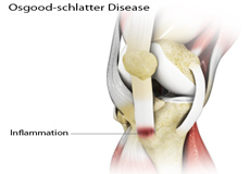
Osgood-Schlatter Disease
Osgood-Schlatter disease refers to an overuse injury that occurs in the knee of growing children and adolescents. This is caused by inflammation of the tendon located below the kneecap (patellar tendon). Children and adolescents who participate in sports such as soccer, gymnastics, basketball, and distance running are at a higher risk of this disease.
Osteochondritis Dissecans of the Knee
Osteochondritis dissecans (OCD) is a problem that affects the knee. The disease behaves much differently in children and for this reason is given a separate name, juvenile osteochondritis dissecans (JOCD).
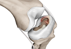
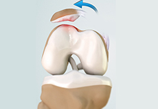
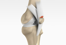
Patellar Instability
Any damage to the supporting ligaments may cause the patella to slip out of the groove either partially (subluxation) or completely (dislocation).
Patellar Tendon Rupture
Patella tendon rupture is the rupture of the tendon that connects the patella (kneecap) to the top portion of the tibia (shinbone).
Patellofemoral Instability
Patellofemoral instability means that the patella (kneecap) moves out of its normal pattern of alignment. This malalignment can damage the underlying soft structures such as muscles and ligaments that hold the knee in place.
Posterolateral Instability
Posterolateral instability, also known as posterolateral rotatory instability (PLRI), is a common pattern of knee instability that results from injuries to the structures that support the outside of the knee joint
Quadriceps Tendon Rupture
The quadriceps can rupture after a fall, direct blow to the leg and when you land on your leg awkwardly from a jump. Quadriceps tendon rupture most commonly occurs in middle-aged people who participate in sports that involve jumping and running.
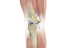
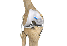
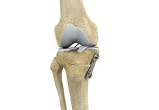
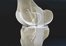
ACL Reconstruction
ACL (anterior cruciate ligament) reconstruction is a commonly performed surgical procedure. With recent advances in arthroscopic surgery, it can now be performed with minimal incision and low complication rates.
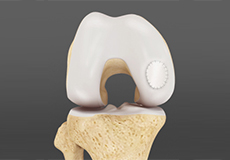
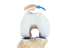
Cartilage Replacement
Cartilage replacement is a surgical procedure performed to replace the worn-out cartilage with new cartilage.
Distal Realignment Procedures
Distal realignment procedures, also known as tibial tubercle transfer (TTT) procedures, are performed to reposition the kneecap after subluxation or dislocation by realigning the tendon under the kneecap to the underlying tibial tubercle.
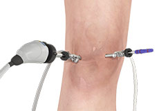
Knee Arthroscopy
Knee arthroscopy is a common surgical procedure performed using an arthroscope, a viewing instrument, to diagnose or treat a knee problem.
Knee Fracture Surgery
A knee fracture is a broken bone or a crack in or around the joint of the knee. This can involve the tibia (shin bone), the kneecap (patella), or femur (thighbone) where they connect with the knee.
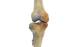
Knee Osteotomy
Knee osteotomy is a surgical procedure in which the upper shinbone (tibia) or lower thighbone (femur) is cut and realigned. It is usually performed in arthritic conditions affecting only one side of your knee.
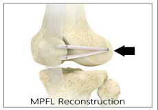
Medial Patellofemoral Ligament Reconstruction
The medial patellofemoral ligament is a band of tissue that extends from the femoral medial epicondyle to the superior aspect of the patella. It is a major ligament that stabilizes the patella and helps in preventing patellar subluxation (partial dislocation) or dislocation.

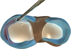
Meniscal Transplantation
Meniscal transplantation is a surgical procedure to replace the damaged meniscus of the knee with healthy cartilage.
Meniscus Replacement
Meniscus replacement is a surgical procedure performed to replace a torn or damaged meniscus in the knee.
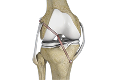
Multiligament Reconstruction of the Knee
Multiligament knee reconstruction is a surgical procedure to repair or replace two or more damaged ligaments of the knee joint. The surgery can be performed using minimally invasive techniques.
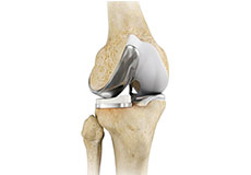
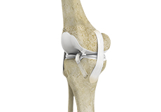
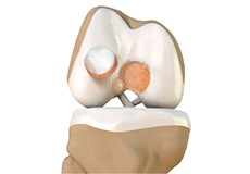
Partial Knee Resurfacing
Partial knee replacement is a surgical procedure which involves resurfacing and replacement of only the diseased surface of the joint instead of the entire joint.
Patellar Tendon Repair
Patellar tendon repair is the surgery performed to reattach the torn tendon to the kneecap and to restore normal function in the affected leg.
Physical Therapy for Knee
Physical therapy is an exercise program that helps you to improve movement, relieve pain, encourage blood flow for faster healing, and restore your physical function and fitness level.
Quadriceps Tendon Repair
Quadriceps tendon is a thick tissue located at the top of the kneecap. The quadriceps tendon works together with the quadriceps muscles to allow us to straighten our leg.
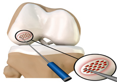
Subchondroplasty
Subchondroplasty is a minimally invasive procedure that is performed to specifically repair chronic BMLs by filling them with a bone substitute material. The bone substitute is then slowly resorbed and replaced with healthy bone, repairing the bone defect. Subchondroplasty also resolves the associated edema. Subchondroplasty may be performed alone or along with other arthroscopic procedures.
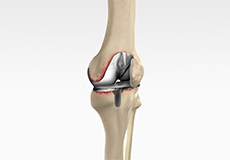
Tibial Tubercle Osteotomy
Tibial tubercle osteotomy is a surgical procedure that is performed along with other procedures to treat patellar instability, patellofemoral pain, and osteoarthritis.
Cartilage Microfracture
Articular or hyaline cartilage is the tissue that covers the bone surface of the knee which helps in smooth interaction between the two bones in the knee joint. It has less capacity to repair by itself because there is no direct blood supply to cartilage.
High Tibial Osteotomy
High tibial osteotomy is a surgical procedure performed to relieve pressure on the damaged site of an arthritic knee joint.
Knee Cartilage Restoration
Knee cartilage restoration is a surgical technique to repair damaged articular cartilage in the knee joint by stimulating new growth of cartilage or by transplanting cartilage into areas with defects in order to relieve pain and restore normal function to the knee.
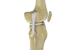
Knee Ligament Reconstruction
Knee ligament reconstruction is a surgical procedure to repair or replace damaged ligaments of the knee joint. The surgery can be performed using minimally invasive techniques..
LCL Reconstruction
LCL reconstruction is a surgical procedure to repair torn or damaged lateral collateral ligament in the knee using a tissue graft taken from another part of the body, or from a donor.
MCL Reconstruction
MCL reconstruction is a minimally invasive surgical procedure in which a tendon graft is utilized to reconstruct the injured MCL.
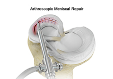
Meniscal Surgery
Meniscal surgery is a surgical procedure employed for the treatment of torn or damaged meniscal tissues in the knee. It is mostly performed as a minimally invasive keyhole procedure.
Meniscectomy
Meniscectomy is a surgical procedure indicated in individuals with torn meniscus where the conservative treatments are a failure to relieve the pain and other symptoms.

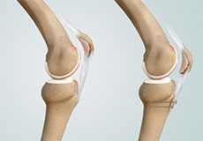
Minimally Invasive Knee Joint Replacement
Total knee replacement is a very successful surgical treatment for knee arthritis. Over the years, minimally invasive knee replacement surgical techniques have been developed to lessen tissue trauma and improve patient outcomes.
Partial Arthroscopic Meniscectomy
Partial arthroscopic meniscectomy is a procedure to remove the damaged part of a meniscus in the knee joint with the help an arthroscope. The meniscus is a C-shaped disc of cartilage between your thighbone and shinbone. There are 2 menisci in each knee.
Partial Meniscectomy
Partial meniscectomy is a surgical procedure to remove the torn portion of the meniscus from the knee joint.
PCL Reconstruction
PCL reconstruction surgery is a procedure to correct torn posterior cruciate ligament (PCL) in the knee using a tissue graft taken from another part of the body, or from a donor.
Posterolateral Corner Reconstruction
Posterolateral corner injury is damage or injury to the structures of the posterolateral corner. The structures of the posterolateral corner include the lateral collateral ligament, the popliteus tendon, and the popliteo-fibular ligament.
Revision Knee Ligament Reconstruction
Revision knee ligament reconstruction is a complex surgical procedure performed to address failures or to correct the undesirable consequences of primary reconstruction surgery on your knee.
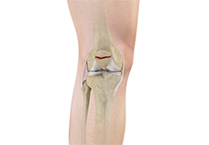
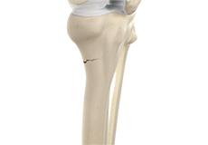
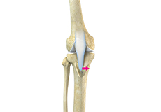
Tibial Eminence Fracture
The tibia or shin bone is a major bone of the leg which connects the knee to the ankle. A fracture or break in the upper part of the tibia is known as a proximal tibial fracture and commonly occurs just below the knee joint.
Tibial Tubercle Transfer
Tibial tubercle transfer is a surgical procedure that is performed along with other procedures to treat patellar instability, patellofemoral pain, and osteoarthritis. The tibial tubercle transfer technique involves realignment of the tibial tubercle (a bump in the front of the shinbone) such that the kneecap (patella) traverses in the center of the femoral groove.
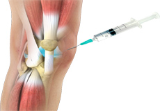
Viscosupplementation
Viscosupplementation refers to the injection of a hyaluronan preparation into the joint. Hyaluronan is a natural substance present in the joint fluid that assists in lubrication. It allows the smooth movement of the cartilage-covered articulating surfaces of the joint.
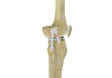
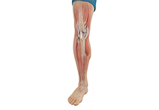
Knee Anatomy
The knee is a complex joint made up of different structures - bones, tendons, ligaments, and muscles. They all work together to maintain the knee’s normal function and provide stability to the knee during movement.
Having a well-functioning healthy knee is essential for our mobility and ability to participate in various activities. Understanding the anatomy of the knee enhances your ability to discuss and choose the right treatment procedure for knee problems with your doctor.
Bones of the Knee
The knee is a hinge joint made up of two bones, the thighbone (femur) and shinbone (tibia). There are two round knobs at the end of the femur called femoral condyles that articulate with the flat surface of the tibia called the tibial plateau. The tibial plateau on the inside of the leg is called the medial tibial plateau and on the outside of the leg, the lateral tibial plateau.
The two femoral condyles form a groove on the front (anterior) side of the knee called the patellofemoral groove. A small bone called the patella sits in this groove and forms the kneecap. It acts as a shield and protects the knee joint from direct trauma.
A fourth bone called the fibula is the other bone of the lower leg. This forms a small joint with the tibia. This joint has very little movement and is not considered a part of the main joint of the knee.
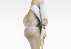
Articular Cartilage and Menisci of the Knee
Movement of the bones causes friction between the articulating surfaces. To reduce this friction, all articulating surfaces involved in the movement are covered with a white, shiny, slippery layer called articular cartilage. The articulating surface of the femoral condyles, tibial plateaus and the back of the patella are covered with this cartilage. The cartilage provides a smooth surface that facilitates easy movement.
To further reduce friction between the articulating surfaces of the bones, the knee joint is lined by a synovial membrane that produces a thick clear fluid called synovial fluid. This fluid lubricates and nourishes the cartilage and bones inside the joint capsule.
Within the knee joint, between the femur and tibia, are two C-shaped cartilaginous structures called menisci. Menisci function to provide stability to the knee by spreading the weight of the upper body across the whole surface of the tibial plateau. The menisci help in load-bearing i.e. it prevents the weight from concentrating onto a small area, which could damage the articular cartilage. The menisci also act as a cushion between the femur and tibia by absorbing the shock produced by activities such as walking, running and jumping.
Ligaments of the Knee
Ligaments are tough bands of tissue that connect one bone to another bone. The ligaments of the knee stabilize the knee joint. There are two important groups of ligaments that hold the bones of the knee joint together, collateral and cruciate ligaments.
Collateral ligaments are present on either side of the knee. They prevent the knee from moving too far during side to side motion. The collateral ligament on the inside is called the medial collateral ligament (MCL) and the collateral ligament on the outside is called the lateral collateral ligament (LCL).
Cruciate ligaments, present inside the knee joint, control the back-and-forth motion of the knee. The cruciate ligament in the front of the knee is called anterior cruciate ligament (ACL) and the cruciate ligament in the back of the knee is called posterior cruciate ligament (PCL).
Muscles of the Knee
There are two major muscles in the knee - the quadriceps and the hamstrings, which enable movement of the knee joint. The quadriceps muscles are located in front of the thigh. When the quadriceps muscles contract, the knee straightens. The hamstrings are located at the back of the thigh. When the hamstring muscles contract, the knee bends.
Tendons of the Knee
A tendon is a tissue that attaches a muscle to a bone. The quadriceps muscles of the knee meet just above the patella and attach to it through a tendon called the quadriceps tendon. The patella further attaches to the tibia through a tendon called the patella tendon. The quadriceps muscle, quadriceps tendon, and patellar tendon all work together to straighten the knee. Similarly, the hamstring muscles at the back of the leg are attached to the knee joint with the hamstring tendon.